Statistically,
almost 30% of the world dies from cancer. Until there’s a miracle drug that cures this disease, surgery is often the best
option. During the surgery, surgeons remove a tumor, but cancer cells can still remain around the margins. Therefore, surgeons biopsy surrounding tissue during surgery and send
it to pathologists for cancer diagnosis. However this process has several limitations such as
sampling error and long preparation time, and it is impossible to biopsy if there are major
nerves and blood vessels in the suspected tissue.
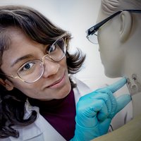
Kyungmin Hwang, CEO of VPIX Medical, Inc.,
conducted research on miniaturization of a confocal microscope into handheld
type (called cCeLL) that can observe tissue microstructure during her doctoral
course at KAIST. In the first year of her Ph.D. program, she founded VPIX
Medical with a neurosurgeon to revolutionize cancer surgery. Her dream is to
see technology that exists only on paper grow into a medical solution
that can truly change the world.
cCeLLTM is a handheld reflective
confocal fluorescence microscope that can acquire unlimited digital tissue
images in real-time but in a non-invasive way. After spraying on or injecting fluorescent
dye into the patient, surgeons can transmit images, instead of actual tissues, to
pathologist, allowing pathologist to provide intraoperative guidance in
real-time so that surgeons can make a surgical decision even before
resection.
VPIX
Medical will give surgeons a tool that lets them work with pathologies in real
time. It finally allows surgeons to share live, high
resolution images from inside patients with pathologists during
surgery. There is no miracle drug to cure cancer, but we believe that we will
soon start making a difference in the world.